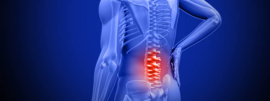
Obtaining an accurate diagnosis that identifies the underlying cause of the pain, and doesn’t just correlate to the symptoms, is important in guiding treatment.
As a foundation of the diagnostic process, the patient provides a detailed description of symptoms and medical history. From this information, a doctor will usually have a general idea of the source of the patient’s pain.
Patient History
Before starting a physical exam, the patient will be asked to provide information regarding symptoms and medical history. Inquiries typically include:
Information about current symptoms: Is the pain better or worse at certain times of day, such as waking up or after work? How far does the pain spread? Are there other symptoms at the same time, such as weakness or numbness? What does the pain feel like—achy, sharp, tight, dull, hot, stinging?
Activity level: Does the person lead a generally more active or sedentary lifestyle? For example, does work require sitting at a desk or standing at an assembly line for long periods of time? How often does the person exercise?
Sleep habits: As a general rule, how many hours of sleep does the patient get? What sleep position is preferred? What kind of and/or quality of mattress and pillow does the patient use?
Posture: What kind of posture feels comfortable or uncomfortable? Does the patient typically sit upright or slouch?
Injuries: Has the person had any recent injuries? Has there been an injury in the past that might be relevant now?
Answers to these questions provide a doctor with a fuller picture of the patient’s daily life, indicating more specific possibilities for low back pain. A medical history is often the most powerful tool for finding a diagnosis.
Physical Exam
The goal of a physical exam is to further narrow down possible causes of pain. A typical physical for low back pain includes some combination of the following steps:
Palpation: A doctor will feel by hand (also called palpation) along the low back to locate any muscle spasms or tightness, areas of tenderness, or joint abnormalities.
Neurologic exam: Diagnosis will likely include a motor exam, which involves a manual movement of the hip, knee and big toe extension and flexion (movement forward and backward) as well as ankle movement. A sensory exam will likely include testing the patient’s reaction to light touch, a pinprick, or other senses in the lower trunk, buttock, and legs.
The range of motion test: The patient may be asked to bend or twist in certain positions. These activities are done to look for positions that worsen or recreate pain, and to see if certain movements are limited by discomfort.
Reflex test: The patient’s reflexes in the legs will be checked to evaluate weakened reflexes and decreased muscle strength. If reflexes are diminished, a nerve root might not be responding, as it should.
Leg raise test: The patient is asked to lie on the back and raise one leg as high and as straight as possible. If this leg raise test recreates low back pain, a herniated disc might be suspected.
Usually, a doctor is able to diagnose low back pain based on the information gleaned from a medical history and a physical exam, and further testing is not needed.
Diagnostic Imaging Tests
An imaging scan is sometimes needed to gain more information on the cause of a patient’s pain. An imaging test may be indicated if the patient’s pain is severe, not relieved within two or three months, and does not get better with nonsurgical treatments.
Common imaging tests include:
X-rays are used to look at the bones of the spine. They show abnormalities, such as arthritis, fractures, bone spurs, or tumors.
A CT scan/Myelogram provides a cross-sectioned image of the spine. In a CT scan (Computed Tomography) an x-ray is sent through the spine, which a computer picks up and reformats into a 3D image. This detailed image allows doctors to look closely at the spine from different angles. Sometimes a myelogram is performed in tandem with a CT scan, in which dye is injected around nerve roots to highlight spinal structures, giving the image more clarity.
An MRI, or Magnetic Resonance Imaging scan, provides a detailed image of spinal structures without using the radiation required with x-rays. An MRI can detect abnormalities with soft tissues, such as muscles, ligaments, and intervertebral discs. An MRI might also be used to locate misalignments or joint overgrowth in the spine.
Injection studies are fluoroscopic-directed injections of local anesthetic and steroid medication into specific anatomic structures. They are helpful in confirming the source of the pain. They are used in diagnosis, in conjunction with rehabilitation, and are considered predictive of surgical outcomes.
Sometimes doctors know what is causing low back pain but not exactly where it is happening, so an imaging test will be used to locate the source more specifically. Imaging tests are also used for patients having surgery so doctors and surgeons can plan the procedure beforehand.
If you are suffering from pain, please contact our office at (516) 419-4480 or (718) 215-1888 to arrange an appointment with our Interventional Pain Management Specialist, Dr. Jeffrey Chacko.


